العوامل التى تؤدى إلى حدوث المرض:
Risk factors for osteoporotic fractures
Risk factors for osteoporotic fractures include female sex, advanced age, low calcium intake, genetic factors, smoking, alcohol abuse, low BMD, low body weight, recurrent falls, personal history of fracture, race or ethnic background, and inadequate physical activity.
Female sex
Menopause occurs approximately at age 51-52 years (range, 42-60 y). Following menopause, levels of circulating estradiol and estrone significantly decrease by around 25% and 75%, respectively.
There is controversy regarding the basic mechanisms underlying the induction of high bone turnover after menopause. Several theories stand out. Direct action of estradiol on osteoclasts has been shown only for avian osteoclasts, but this mechanism remains a clear favorite. Bone resorption is the unique function of the osteoclast. In the avian model, estradiol decreases the development and activity of osteoclasts and increases the activity of osteoblasts directly. Estrogen deficiency induces increased generation and activity of osteoclasts, which perforate bone trabeculae, reducing their strength and increasing fracture risk. The life span of functional osteoclasts and thus the amount of bone that osteoclasts resorbed may also be enhanced following estrogen deficiency.
Estrogen may affect osteoclast function by promoting apoptosis. It has been shown that 17beta-estradiol promotes apoptosis of murine osteoclasts in vitro and in vivo by 2-3 times. This suggests that estrogen may prevent excessive bone loss before and after the menopause by limiting the life span of osteoclasts.
Recently, estrogen has also been shown to regulate secretion of osteoprotegerin, an inhibitor of osteoclast differentiation.
Most of the estriol present in the circulation after menopause represents the extraendocrine conversion of androgen precursors in muscle and adipose tissue to estriol. This conversion in adipose tissue may explain why obese patients are relatively protected against osteoporosis and fractures compared with asthenic individuals.
Women undergoing early menopause or oophorectomy have accelerated bone loss and a higher incidence of fractures. Amenorrhea also predisposes women to osteoporosis. Those experiencing early menopause usually have prolonged periods of oligomenorrhea, a trait that has a strong genetic predisposition. Thus, these patients have repeat periods of increased bone loss and low bone mass.
Early and late estrogen deficiency probably affects bone mass by means of different mechanisms. Early estrogen deficiency, ie, that occurring before age 25 years when patients attain peak bone mass, probably affects bone maturation and formation during bone modeling. This leads to a thinner and a more slender skeleton. Early estrogen deficiency occurs in Turner syndrome, hyperprolactinemic amenorrhea, and amenorrhea among athletes. By contrast, normal menopause and late estrogen deficiency (eg, that following oophorectomy) induces a state of accelerated bone loss from increased osteon activation frequency. Recent work has also demonstrated increased cellular sensitivity to parathormone (PTH) in patients with osteoporosis.
Advanced age
Bone mass peaks at age 25 years. Thereafter, the bone mass in both sexes remains stable until age 45-55 years, when accelerated bone loss ensues in women and a more gradual loss commences in men. The accelerated bone loss in women causes the loss of 25-30% of skeletal mass over 5-10 years, followed by a slower phase with stable loss rates of 0.5-1% per year. Males do not have an accelerated bone loss, but rather, a stable loss rate.
Recent studies suggest that both sexes undergo a late phase of accelerated bone loss in old age. The mechanisms by which bone loss occurs after age 35 years are poorly understood, but several factors related to age-dependent changes in skeletal and calcium homeostasis have been implicated; these include estrogen deficiency, reduced osteoprotegerin levels, reduced calcium and vitamin D intake, impaired calcium and vitamin D absorption, increased interleukin-1 and interleukin-6 levels, tumor necrosis factor-alpha, increased bone resorption and turnover, impaired osteoblast function, decreased insulin-like growth factor secretion, decreased transforming growth factor–beta secretion, and reduced core-binding factor–1 levels.
Recent work has shown that in both males and females the effects of estrogen deficiency on the rate of bone loss last throughout life in both sexes. In males, the bone loss rate with increasing age is also related to circulating estradiol levels.
Low calcium intake
Calcium is an essential mineral in maintaining nerve function, muscle function, and bone mineralization, and it is involved in the control of several intracellular processes. Physiologically, several hormonal systems work to maintain calcium homeostasis. Vitamin D is essential for the absorption of calcium from the gut. Calcium is then transported via the blood to bone, where it is incorporated in the bone matrix during calcification. During periods of calcium deficiency from decreased intake or decreased absorption, bone acts as a buffer, maintaining a constant level of calcium in the blood.
Calcium can be removed from bone either through transport over the osteocyte-lining cell system, which is responsible for the rapid regulation of serum calcium, or via the liberation from the bone matrix through osteoclastic resorption. Calcium loss also occurs through the gut, kidney, and skin. The kidney plays an important role in calcium homeostasis by affecting PTH levels.
Adequate calcium intake is important to maintain normal calcium homeostasis and to protect the bones from excessive calcium loss. If calcium intake is low, mechanisms that increase secretion of PTH are brought into play, resulting in a high-turnover state and possible negative effects on bone mass. The minimum calcium intake necessary to maintain skeletal health is difficult to define. Nutrition may affect peak bone mass.
Matkovic et al compared incidence of femoral neck fractures in people living in 2 geographically and dietetically separated valleys in the area formerly known as Yugoslavia. They found a reduced incidence of femoral neck fractures among individuals living in the valley with the higher calcium intake. The difference is probably attributable to differences in peak bone mass.
The impact that calcium has on developing and maintaining bone mass varies throughout life. To reduce the risk of osteoporosis, calcium intake should be highest during adolescence, pregnancy, and old age.
Genetic factors
About 60% of a person's peak bone mass is genetically determined. A woman whose mother has osteoporosis is more likely to have the condition. The remaining 40% of one's peak bone mass is attributed to dietary factors, physical activity, medication use, and lifestyle.
Smoking
Smoking is an important risk factor for osteoporosis. Several epidemiologic studies and a recent meta-analysis showed a significant impact of smoking on bone mass, especially in older age groups. However, 2 large-scale European studies did not show any significant effect on osteoporotic fractures.
Smokers are known to experience menopause earlier than nonsmokers, and because they are slimmer than nonsmokers, they have reduced extraendocrine production of estrogens, as in adipose tissue. Smokers may also have increased metabolic clearance rate of estrogens. In addition, smoking may directly inhibit osteoblast function.
Alcohol
Previous or present alcoholism is a risk factor for the development of osteoporosis. Moreover, inebriation increases the risk of falls and thus potentiates fractures. Alcohol affects osteoblast proliferation in vitro and reduces matrix protein synthesis in vivo. It exerts a direct toxic effect on other bone cells as well. Even so, 2 large European studies showed no significant effect of moderate alcohol consumption on osteoporotic fracture risk in women.
Hormones
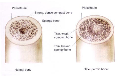
Bone remodeling is responsible for the replacement of old bone with new. This process initiated by osteoclastic activity responsible for the resorption of old bone. Bone resorption lasts for 20-40 days and is followed by osteoblast formation of unmineralized bone matrix, which subsequently mineralizes over the next 100-150 days. Under physiologic conditions, homeostasis occurs between bone resorption and bone formation. However, during pathologic conditions, negative bone balance may occur. Occasionally, positive balance can lead to the overproduction of bone.
Calcium homeostasis is maintained through a complex interaction between the parathyroid glands, skin, gut, and kidneys. In this process, serum calcium levels are maintained within a narrow physiologic range. Normally, a negative feedback loop involving PTH and 1,25-dihydroxy vitamin D-3, or 1,25-(OH)2D3, maintains body calcium levels despite large variations in the influx and efflux of calcium from the body. A negative feedback loop also exists between serum calcium and PTH to inhibit secretion of the latter.
Renal parenchymal disease causes low levels of 1,25-(OH)2D3 resulting in compensatory hyperparathyroidism, which increases bone resorption and bone turnover. While bone loss in early menopause is mainly related to decreased endogenous estrogen production, bone loss after age 65 years involves mechanisms more closely related to disturbance of calcium homeostasis due to reduced vitamin D and calcium intake.
With aging, calcium intake is reduced. Production of active vitamin D in the skin is also decreased resulting in decreased absorption of consumed calcium. Reduced calcium absorption may cause secondary hyperparathyroidism, which in turn accelerates bone loss through increased osteoclast activity and hence bone turnover.
Impaired osteoblast function, however, causes accelerated bone loss. Like fibroblasts, osteoblasts undergo cellular aging with increasing age. As a result, collagen matrix synthesis and secretion of other osteotropic factors decrease. This leads to lower rates of bone formation in the elderly. The main difference between osteoporotic women and non-osteoporotic women is defective bone formation. Osteoporotic women without fractures have significantly thinner bone structural units compared with age-matched controls. Genetic and hormonal factors besides aging may also contribute to osteoblastic insufficiency.
Estrogens increase serum levels of 1,25-(OH)2D3. In osteoporotic women, calcium absorption increases with calcitriol supplementation. This effect has been considered one of the indirect effects of estradiol, and it may explain the beneficial role of estrogen supplementation in the prophylaxis against osteoporotic fractures.
Low body weight
Body weight and rates of hip fracture are inversely related. In the Framingham study, the relative risk of fracture was 0.63 in individuals who were 114-123% overweight and 0.33 in individuals more than 138% overweight. Obesity appears to protect the skeleton in several ways: by increased the production of estrone in fatty tissue, by improving vitamin D storage in fatty tissues, by exerting a cushioning effect in association with falls, and by creating a larger skeleton as a result of increased weight bearing.
Recurrent falls
Both falls and reduced skeletal resistance are important determinants of fracture risk. The risk of falls increases exponentially after age 40 years and is greater in women than in men. Most falls that lead to fractures, especially age-related fractures, occur from a standing height or shorter distance. Most age-related fractures are associated with slips, trips, and drop attacks. Such falls cause the majority of fractures of the distal radius and a substantial proportion of hip fractures. Falls down stairs are the major cause of vertebral fractures associated with spinal cord injuries. Naturally, falls from heights are an important but less common cause of fractures.
Preventing falls is important prophylaxis against osteoporotic fractures. Predisposing factors, such as postural hypotension or drowsiness due to drug use, should be detected and treated. If necessary, patients should receive physiotherapy and walking aids to improve their balance and righting reflexes.
Personal history of fracture
Lindsay and colleagues determined the incidence of recurrent vertebral fractures in women receiving placebo in 4 large, 3-year clinical trials to evaluate the efficacy of bisphosphonates for treatment of postmenopausal osteoporosis. The cumulative incidence of new vertebral fractures in the first year was 6.6%. Among women who had an incident vertebral fracture, the incidence of another vertebral fracture in the subsequent year was 19.2%. The presence of a vertebral fracture at study baseline was associated with an increased risk of another fracture.
Race or ethnic background
Racial differences in peak BMD partly may account for racial differences in the incidence of osteoporosis and fractures. Populations of African origin have higher bone mass and lower rates of fractures, as compared with white populations. BMD is greater in adult blacks than in whites. Also, prepubertal BMD in the hip, trochanter, and femoral neck is higher in black males than in white males. Reduced thickness of the femoral neck and shaft cortex, a wider intertrochanteric region, and a longer hip-axis length are thought to contribute to the higher incidence of hip fracture among white women. In comparison, women of African origin on average have thicker cortical bone in the hip, a shorter hip-axis length, and smaller intertrochanteric widths.
Although Asian women have lower bone mass than that of Caucasian women, they have a lower rate of hip fractures. Several postulates have been forwarded to explain this discrepancy, including a shorter hip-axis length in the Asian women, higher activity levels in childhood, and, the cultural practice of taking care of the elderly, and the practice in which women are not allowed to leave their beds (which reduces the opportunity for falling). Hispanic women tend to have bone density equivalent to that of white women, but they have one half as many fractures. This probably related to cultural differences or possible may be related to the microarchitecture of the bone itself.
There are major differences between BMD values in European population samples, which, with variations in anthropometric variables, have the potential to contribute substantially to variations in rates of osteoporotic fracture risk. The highest rates are in Scandinavian countries, likely secondary to reduced sun exposure and hence less vitamin D formation.
Inadequate physical activity
Physical activity is essential for bone remodeling. The skeleton needs continuous physical stimulation to maintain healthy bones, otherwise bone loss ensues.
Osteoblast activity is sensitive to mechanical stresses. Experiments of repetitive physical stress on bone have shown profound increases in bone formation in stressed bone. Significant bone loss occurs from immobilization or during space flight. Studies have shown that physically active women have a higher bone mineral content than women who are less active. Antigravity exercises, such as dancing or running, seem to be more effective than swimming in maintaining BMD. In vertebrae, the preferential loss of horizontal trabeculae leads to compensatory thickening of vertical trabeculae. The correction of tooth alignment exploits physical stress to create changes in bone remodeling in the jaw.
The steady decrease in general physical activity in the population is probably one of the factors responsible for the increasing prevalence of osteoporosis over the last 10 years. Several studies in perimenopausal women have shown increases in bone mass between 5-7% over a 3-year period following the institution of an exercise regimen compared with sedentary controls. Therefore, a reasonable amount of physical activity throughout life may protect individuals against bone loss.
It is unlikely that physical activity alone can offset the 30-40% loss of bone occurring after menopause. In fact, a meta-analysis of all controlled clinical trials showed no significant effects of physical activity on bone mass. Further, long-term clinical trials have shown no fracture protection from exercise. To the contrary, one study showed an increased fracture risk in a population of older women who walked for exercise.
Prolonged heavy exercise may have deleterious effects on bone mass. Extremely high levels of physical activity in young women may produce hypothalamic amenorrhea and hence estrogen deficiency.
Involutional osteoporosis in men
Data suggest that hypogonadism is but one determinant of male osteoporosis. In recent publications about male osteoporosis, only 12% of men had low s-testosterone levels. Other research has shown that male estrogen deficiency may also be an important cause of male osteoporosis. Low bone mass in men may be related to aromatase deficiency, which normally converts testosterone to estradiol.
المرجع:
http://www.e-radiography.net/radpath/o/osteoporosis.htm